The Dynamic Dance of Fluids and Lasers: Rethinking Dentinal Sensitivity
The intricate interaction between fluid kinetics and innovative technologies is reshaping our understanding of discomfort in specific dental conditions. Modern advancements showcase groundbreaking strategies, surpassing traditional approaches, offering hope for long-lasting relief. The merging of legacy theories with contemporary methods signals a promising shift in addressing these challenges effectively.
Unlocking the Microscopic Hydraulic System
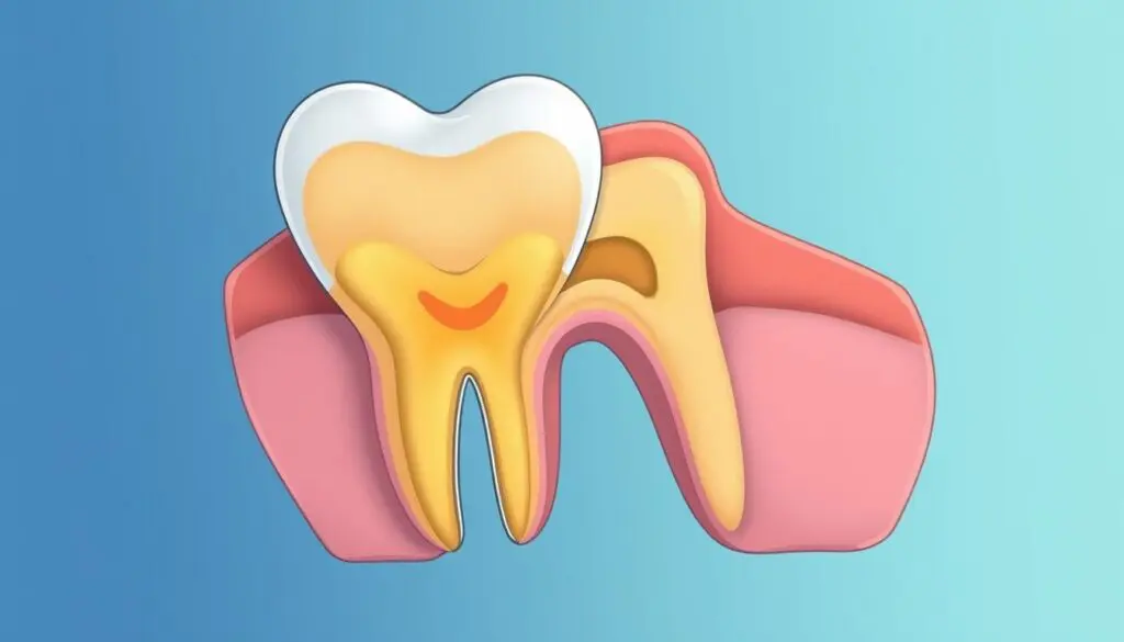
The Hidden Dynamic World Inside Your Teeth
To the naked eye, a tooth appears to be a solid, static object, much like a polished stone or a piece of ivory. However, if we were to zoom in at a microscopic level, the reality is drastically different. The layer beneath the enamel, known as dentin, is not a solid block but rather a complex, porous structure. It is permeated by thousands of microscopic channels radiating outward from the nerve center. These channels are not empty; they are filled with a specialized biological fluid. This means that our teeth are not just calcified tools for chewing but contain a dynamic hydraulic system that obeys the same laws of physics found in nature.
When we discuss fluid mechanics, we often think of large-scale examples like water rushing through a dam or air flowing over an airplane wing. Surprisingly, these same physical principles apply within the microscopic confines of your mouth. The fluid inside these tiny channels is constantly in a state of potential motion. It is incredibly sensitive to changes in the external environment. This fluid does not simply sit stagnant; it expands, contracts, and shifts position based on what happens on the tooth's surface. Understanding this behavior is crucial because it transforms our view of dental pain from a mysterious ailment into a logical physical phenomenon.
For decades, dental science struggled to explain exactly how sensation traveled through the hard tissue of the tooth to the nerve, given that the nerves themselves do not extend all the way to the surface. The answer lies in the movement of this fluid. When the protective outer layers of the tooth are compromised—due to gum recession or enamel wear—these fluid-filled channels become exposed to the outside world. This exposure turns the hydraulic system into a direct transmission line for sensory information. By applying concepts like pressure gradients and flow velocity to biological tissue, we can begin to understand why a sip of cold water or a breath of winter air translates into a sharp, sudden jolt.
From Physical Stimulus to Sharp Sensation
The Chain Reaction of Temperature and Pressure
The prevailing scientific explanation for this sensitivity revolves on the concept that fluid movement acts as a mechanical trigger. When the dentin is exposed, stimuli do not need to touch the nerve directly to cause pain. Instead, they disrupt the equilibrium of the fluid within the channels. Imagine the fluid acting like a piston inside a cylinder. When an external force is applied, the "piston" moves, and this physical displacement is what eventually rings the alarm bells in the tooth's nerve center. The speed and direction of this movement are critical factors in the intensity of the sensation experienced.
Different types of everyday stimuli cause the fluid to move in specific ways, creating a "hydrodynamic dance" within the tooth. For instance, when you eat ice cream or inhale cold air, the temperature drop causes the fluid volume to contract rapidly. This creates an outward suction force, pulling fluid away from the nerve. Conversely, hot foods can cause expansion, pushing fluid inward. Sweet or salty foods introduce a change in osmotic pressure, drawing fluid out toward the surface. Even the blast of air used by a dentist to dry a tooth causes rapid evaporation, which pulls fluid outward to replace what was lost.
| Stimulus Type | Physical Reaction in Micro-Channels | Direction of Fluid Force |
|---|---|---|
| Cold Temperatures | Rapid contraction of fluid volume | Outward (Suction) |
| Hot Temperatures | Expansion of fluid volume | Inward (Pressure) |
| Dehydration / Air Blast | Evaporation causing capillary flow | Rapid Outward Flow |
| Sugary / Salty Foods | Change in osmotic pressure gradients | Outward (Osmotic pull) |
| Tactile / Touch | Direct mechanical displacement | Variable / Inward |
Regardless of whether the trigger is thermal, chemical, or mechanical, the result is a rapid shift in the fluid column. Research suggests that this fluid can move at speeds of several millimeters per second—a veritable rushing river at the microscopic scale. This rapid flow exerts a "shear stress" on the nerve endings and specialized cells located at the pulp-dentin border. These cells are equipped with mechanical receptors that detect distortion. When the fluid rushes past, it physically deforms these receptors, converting the mechanical energy into an electrical signal that the brain interprets as sharp pain.
Why Flow Velocity Matters
The relationship between the speed of the fluid and the pain perceived is direct and significant. A slow, gradual movement might not trigger a response, but a sudden acceleration creates a high-impact event for the nerve. This explains why the pain is often sharp and shooting rather than dull and throbbing. The system is designed to detect rapid changes, likely as a defense mechanism to prevent further damage to the tooth structure.
Modern imaging and fluid dynamics studies have confirmed that the diameter of the channels plays a massive role in this sensitivity. The wider the channel, the easier it is for fluid to flow, and the faster it can move. This is consistent with the observation that as people age, their channels often naturally narrow or become blocked with mineral deposits, leading to a reduction in sensitivity even if gum recession is present. This natural blocking process reduces the volume of fluid that can be displaced, effectively dampening the hydraulic signal before it can reach the nerve.
This understanding has fundamentally shifted how we approach treatment. The goal is no longer just to numb the nerve with anesthetics, which is a temporary fix. Instead, the objective is to stop the fluid from moving in the first place. If we can physically block the opening of the channel or reduce the diameter of the tunnel, we can prevent the hydraulic shift from occurring. This is the logic behind most modern desensitizing therapies: they are essentially attempting to put a lid on the detailed hydraulic machinery operating inside the tooth.
Advanced Interventions: The Era of Light Energy
High-Tech Barriers and Cellular Biostimulation
While traditional toothpastes and varnishes aim to deposit particles into the open channels to clog them, these methods often provide only temporary relief. The deposited layers can be brushed away or dissolved by acidic foods over time. This limitation has paved the way for the use of high-energy light technologies in managing sensitivity. The application of laser energy offers a fundamentally different approach: instead of simply placing a cork in the bottle, lasers can alter the structure of the bottle itself to seal it shut.
When specific wavelengths of light energy are directed at the exposed dentin, the interaction creates a localized thermal effect. This heat can melt the mineral components of the tooth surface and the proteins within the fluid. As these materials cool, they re-crystallize and fuse together, creating a non-porous, glass-like surface. This process effectively "welds" the openings of the microscopic channels shut. Because this is a structural change rather than a superficial coating, the seal is far more durable and resistant to the daily wear and tear of brushing and eating.
| Feature | Traditional Desensitizing Agents | Laser-Assisted Treatment |
|---|---|---|
| Mechanism of Action | Mechanical clogging (precipitation) | Melting, fusion, and protein coagulation |
| Durability | Temporary; requires daily re-application | Long-lasting; structural alteration |
| Immediate Effect | Gradual build-up over weeks | Often immediate relief after procedure |
| Resistance to Acid | Low; easily dissolved by diet | High; creates resistant surface layer |
| Clinical Setting | At-home (toothpaste) or simple varnish | Professional in-office procedure |
Beyond the physical sealing of the tubes, certain light therapies offer a biological benefit known as biostimulation. Low-level energy can be used to sedate the hyperactive nerves directly. By targeting the cellular mitochondria, this gentle energy promotes healing and raises the pain threshold of the nerve endings. This dual approach—physically blocking the hydraulic trigger while simultaneously calming the biological alarm system—represents the cutting edge of comfort management. It moves the treatment from simple symptom masking to a comprehensive restoration of function, allowing patients to enjoy cold drinks and sweet treats without the fear of that familiar, sharp jolt.
Q&A
-
What are exposed dentinal tubules and how do they affect dental health?
Exposed dentinal tubules occur when the protective enamel and cementum layers of a tooth are worn away, revealing the underlying dentin. This exposure can lead to increased sensitivity and discomfort, as the tubules contain fluid and nerve endings that react to temperature changes and pressure.
-
How does fluid movement within dentinal tubules relate to Brännström's Theory?
Brännström's Theory, also known as the hydrodynamic theory, suggests that the movement of fluid within dentinal tubules is responsible for the sensation of pain and sensitivity in teeth. When stimuli such as cold or pressure cause the fluid to move, it triggers nerve responses that the brain interprets as pain.
-
What role do desensitizing agents play in managing dental sensitivity?
Desensitizing agents are used to alleviate tooth sensitivity by either blocking the nerve endings in the tubules or by forming a barrier over the exposed dentin. Common agents include potassium nitrate, fluoride, and strontium chloride, which help reduce fluid movement and desensitize the nerves.
-
How can cervical abrasion lead to exposed dentinal tubules, and what are the common causes?
Cervical abrasion is the wear of the tooth structure near the gum line, often caused by aggressive brushing, acidic diets, or bruxism (teeth grinding). This abrasion can expose dentinal tubules, leading to increased sensitivity and potential for decay if not addressed.
-
In what ways can laser treatment be used to treat exposed dentinal tubules?
Laser treatment is a modern approach to treating exposed dentinal tubules. It works by sealing the tubules, reducing fluid movement, and thus decreasing sensitivity. This method is minimally invasive and can provide long-lasting relief from discomfort associated with dentin exposure.