Hidden Battles: The Chronic Dance Between Periapical Abscess and Bone Resorption
In the shadowy interplay of dental ailments, a persistent struggle unfolds deep within the oral cavity. Often underestimated, this struggle manifests as an insidious aggressor, fostering an environment ripe for discomfort and chronic progression. Its menacing advance reshapes oral landscapes, presenting visible signs of disturbance, complexity, and resilience.
The Invisible Erosion of the Jaw's Foundation
When Defense Becomes Destruction
The trouble brewing at the root of a tooth is rarely just about swelling or localized pain; it is a complex biological standoff. When bacteria breach the inner sanctum of a tooth, the body deploys a sophisticated defense system to contain the invasion. However, the jawbone surrounding the root tip becomes the primary battlefield for this confrontation. Interestingly, the loss of bone density often observed in these cases is not primarily caused by the bacteria eating away at the structure. Instead, it is a byproduct of the body’s own immune response, a phenomenon scientifically referred to as bone resorption.
To fight off the infection, the immune system recruits specialized cells to the site. To create physical space for these defense cells to operate and to isolate the infection, the body initiates a process that dissolves the surrounding bone. Under normal circumstances, our skeletal system maintains a harmony between cells that build bone and cells that break it down. However, in the presence of chronic inflammation—often diagnosed as apical periodontitis—this balance is disrupted. The inflammatory markers trigger an overactivity of the bone-destroying cells. Consequently, the jawbone is sacrificed in a desperate attempt to prevent the bacteria from spreading deeper into the body. This "friendly fire" is a silent process, eroding the foundation of the tooth without immediate, sharp pain.
Deciphering the Shadows on the Screen
This internal erosion leaves behind clues that are invisible to the naked eye but glaringly obvious to a trained professional equipped with imaging technology. On a standard dental X-ray, these areas of bone loss appear as dark, shadowy halos around the root tip. This visual characteristic, known as radiolucency, indicates that the hard, mineralized bone has been replaced by softer inflammatory tissue or a void. It serves as a historical map of the battle that has been raging silently beneath the gum line.
Interpreting these shadows is akin to solving a high-stakes puzzle. A dark spot could represent a fluid-filled cyst, a granuloma (a mass of granulation tissue), or an acute infection. Because two-dimensional X-rays can sometimes be misleading—flattening complex structures onto a single plane—modern dentistry often utilizes 3D imaging (CBCT) to gauge the true extent of the damage. These advanced images can reveal if the lesion has perforated the cortical plate of the jaw or wrapped around complex root anatomy. Identifying the precise nature of this radiolucency is critical, as it dictates whether the tooth can be saved through conservative treatment or if more invasive measures are required.
| Feature Comparison | 2D X-Ray (Standard) | 3D CBCT Imaging |
|---|---|---|
| Visual Depth | Flat, single-plane view; structures may overlap. | Volumetric view; allows rotation and slicing of images. |
| Lesion Detection | Good for general screening; may miss early bone changes. | Highly sensitive; reveals the exact size and location of the void. |
| Anatomical Context | Shows general relationship to neighboring teeth. | Shows proximity to nerves, sinuses, and cortical bone boundaries. |
| Primary Use Case | Routine check-ups and initial diagnosis. | Complex surgical planning and evaluating difficult root canals. |
The Pressure Cooker and the Escape Route
The Mechanics of Agony and Fluid Buildup
Before the body manages to erode enough bone to create space, the initial phase of infection is often marked by intense, throbbing pressure. This occurs because the root tip is encased in a rigid environment of tooth and bone. When the immune system clashes with bacteria, the byproduct is purulent exudate, commonly known as pus—a mixture of dead white blood cells, bacteria, and tissue debris. In the confined space of the jawbone, there is nowhere for this fluid to go.
This situation creates a hydraulic pressure trap, similar to over-inflating a balloon inside a stone box. The rapid increase in internal pressure compresses the nerve endings in the surrounding area, leading to the excruciating, pulsating pain often associated with acute dental infections. This pain is a biological alarm system, screaming that the containment breach is critical. Patients often report that the tooth feels "high" when biting down or is incredibly sensitive to touch. This is not merely a symptom of a cavity but a sign that the accumulation of fluids has reached a critical mass within the bony housing.
The Tunnel to the Surface
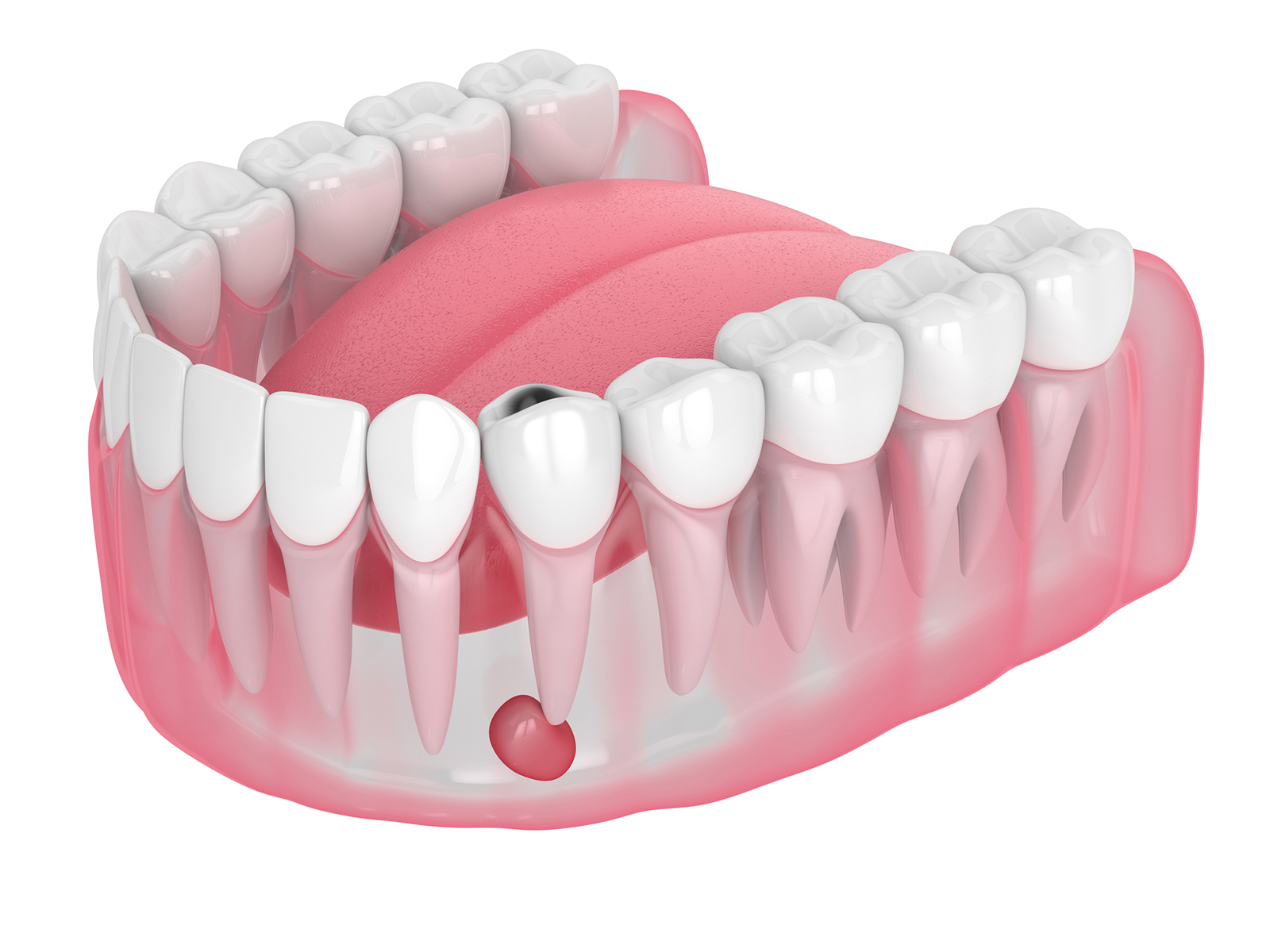
If the pressure is not relieved through dental intervention, the fluid will eventually seek the path of least resistance to escape. The corrosive nature of the inflammation softens the bone, allowing the infection to tunnel through the jaw until it breaches the gum tissue. This process results in fistula formation, a biological drainage tract that connects the abscess at the root to the oral cavity.
Clinically, this often presents as a small, pimple-like bump on the gums, sometimes accompanied by a discharge that leaves a foul taste in the mouth. When this tunnel opens and the fluid drains, the internal pressure drops significantly, and the intense pain often vanishes instantly. This creates a dangerous illusion of healing. Patients may believe the problem has resolved because the pain is gone, but the reality is quite the opposite. The infection has merely transitioned from an acute, contained phase to a chronic, open state. The destruction of bone continues unabated, now with a permanent exit route, potentially compromising the long-term retention of the tooth and systemic health.
Interventions to Halt the Cycle
Releasing the Hydraulic Trap
The cornerstone of treating these deep-seated infections is establishing drainage. Without physically removing the buildup of bacteria and toxins, the body cannot effectively heal the damaged bone. Drainage procedures are designed to release the pressure and remove the source of infection. The most common method is initiating root canal therapy, where the dentist accesses the inner chamber of the tooth to clean out the infected pulp and allow the accumulated pus to drain through the tooth itself.
In cases where the infection has spread significantly or a large swelling has formed, an incision into the gum tissue might be necessary to facilitate immediate relief. By mechanically removing the irritants and disinfecting the complex canal system, the dentist changes the environment from one that favors bacterial growth to one that promotes healing. Once the continuous stream of toxins is halted, the overactive bone-destroying cells calm down, and the body’s bone-building cells can begin the slow work of repairing the damage.
The Critical Importance of Timely Action
Ignoring the signs of deep dental infection—whether it’s the shadow on an X-ray or the pimple on the gum—can lead to severe consequences. The "wait and see" approach is particularly risky because the absence of pain does not equal the absence of disease. A chronic abscess acts as a reservoir of bacteria that can periodically flare up or spread to other parts of the head and neck.
Effective management requires a shift in perspective: from treating symptoms (pain) to treating the biological cause (infection). Modern endodontic techniques aim to save the natural tooth whenever possible, but success relies heavily on how much bone structure remains intact. The earlier the intervention, the higher the likelihood that the radiolucency will heal and the bone will regenerate. Understanding that the silence after the pain is merely a truce, not a victory, is essential for maintaining oral health.
Q&A
-
What is fistula formation and how is it related to dental conditions?
Fistula formation is a pathological channel that connects an internal body cavity to the skin surface or another cavity. In dental conditions, it often occurs as a result of chronic infection, such as apical periodontitis, where pus from the infection creates a tract through bone and soft tissue to drain externally.
-
How does apical periodontitis lead to bone resorption?
Apical periodontitis is an inflammatory condition affecting the apex of a tooth's root, typically resulting from bacterial infection in the root canal system. The body's immune response to the infection can cause the resorption of surrounding bone tissue as it attempts to eliminate the infection, leading to loss of bone density and structural integrity.
-
What role does purulent exudate play in dental infections?
Purulent exudate is a thick, yellowish fluid composed of white blood cells, dead tissue, and bacteria, commonly referred to as pus. In dental infections, such as those associated with apical periodontitis, purulent exudate indicates the presence of an active infection and can contribute to the formation of fistulas or sinus tracts as the body attempts to drain the infection.
-
Why is radiolucency significant in diagnosing dental issues?
Radiolucency on dental X-rays indicates areas where bone density is reduced, often due to conditions like bone resorption associated with apical periodontitis. Identifying radiolucent areas helps dentists diagnose the extent of the infection and assess the severity of bone loss, guiding treatment decisions.
-
What are drainage procedures and how are they used in managing dental infections?
Drainage procedures are medical interventions used to evacuate pus or fluid from an infected area, alleviating pressure and pain. In dentistry, they are often employed to treat abscesses or fistulas by creating an incision or channel that allows purulent exudate to exit, thereby reducing infection and facilitating healing. These procedures are typically accompanied by antibiotic therapy and sometimes surgical intervention to address the root cause of the infection.